Clinical Detection The value of Acanthocyte in identifying the source of hematuria
Urinalyses, as one of the three widely used routine tests in clinical practice, has important value in screening, distinguishing, and observing the efficacy of urinary systemic diseases. Hematuria is a dangerous signal, therefore, early diagnosis of the underlying disease is necessary for patients with hematuria. The diagnosis of hematuria first needs to distinguish whether it is glomerular hematuria or non glomerular hematuria. Glomerular hematuria is commonly seen in various primary or secondary glomerulonephritis, while non glomerulonephritis is commonly seen in kidney stones, kidney tumors, etc. The identification of the source of hematuria can be based on the morphology of red blood cells, so the identification of red blood cell size, morphology, and subdivision is particularly important, especially for Acanthocyte, which is landmark red blood cell in renal hematuria.
Case:
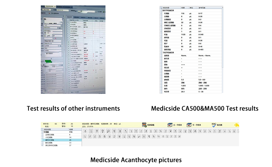
Patient Ms. Deng, 47 years old, visited the hospital on July 2, 2023. The urinalyses results are shown in the following figure:

On the outpatient testing instrument, the test results showed dry chemical urine protein+urine occult blood 3+, and the total number of formed red blood cells was only 99.8/ul, indicating mixed hematuria. Due to the inability of the instrument to perform detailed classification of red blood cells and review of red blood cell images, the samples were tested on the Medicside CA-500&MA-500 automatic urine assembly line. The report showed dry chemical urine protein+urine occult blood 3+, and a total of 116 formed red blood cell test results were obtained. The morphology of red blood cells was further classified: 4 normal red blood cells/ul, 14 small red blood cells/ul, 28 Acanthocyte/ul, and 33 Erythrocyte Ghost/ul, Other poikilocytes 37/ul. Through the Medicside detection, we can see that red blood cells are mainly abnormal in morphology and exhibit two or more multiple morphological changes. Among them, Acanthocyte accounts for 23.85% of the total number of red blood cells, indicating glomerular hematuria. The diagnosis of the patient is IGA nephropathy (a group of glomerular diseases characterized by mesangial hyperplasia and significantly diffuse IgA deposition in the mesangial area. Its clinical manifestations are diverse, with hematuria being the most common). Therefore, the analysis and recognition of the size and morphology of red blood cells is an important basis for distinguishing the source of hematuria, and is of great significance for the diagnosis of clinical diseases.
Acanthocyte introduction:
In July 2021, CHINESE JOURNAL OF LABORATORY MEDICINE published Expert consensus on name and result report of urine formed elements, this consensus provides a systematic and detailed naming and refinement of the formed components in urine, providing a standardized theoretical basis for morphological examination personnel. There is a detailed introduction to Acethocyte:
Acanthocyte is a type of heteromorphic red blood cells, also known as G cells, commonly found in glomerular diseases and also known as glomerular red blood cells. It is a landmark red blood cell in renal hematuria.
The source and mechanism of Acanthocyte: Red blood cells undergo mechanical damage caused by strong stretching or compression when passing through the pathological glomerular basement membrane, resulting in morphological changes in renal tubular filtrate affected by continuous changes in pH and osmotic pressure.
Acanthocyte morphology: Red blood cells vary in size, with one or more spiny protrusions of varying sizes at the edge or center of the cell, or pseudopodia, resembling spores; The center is shaped like a mouth, target, or irregular shape.
Classification of Acanthocyte: Scholars once classified G cells into five categories: G1, G2, G3, G4, and G5. In terms of morphology, G1 cells are red blood cells with more than one spore like protrusion and obvious hemoglobin overflow. After hemoglobin overflow, a faint shadow with spores is formed; G2 cells are spiny shaped, with spherical shapes, varying sizes, and thick membranes. The concentration of hemoglobin in the cells is uneven, and there are multiple spiny protrusions on the surface of the cells; G3 cells are doughnut like red blood cells with irregular convex and concave surfaces; G4 cells are tumor like protrusions or sprouting red blood cells; G5 cells are significantly reduced red blood cells, appearing as small rings due to reduced refraction. In recent years, it has been found that not all G cells are glomerular derived, as G4 cells can be seen in non glomerular hematuria. In addition, there is overlap between the morphological descriptions of different types of G cells and the standardized naming of red blood cells, which can easily cause confusion in clinical work. Therefore, the use of G cell classification is no longer recommended. It is recommended to classify G1 and G2 cells as spinous cells, G3 cells as circular red blood cells, G4 cells as Temp1, and G5 cells as serrated red blood cells based on their morphological characteristics.
The value of spinous cells in identifying the source of hematuria: Due to the classification of G1 and G2 cells as spinous cells, it is currently widely believed that G1 cells ≥ 5% are characteristic markers of glomerular hematuria. However, in clinical practice, it is relatively difficult to distinguish between G1 and G2 cells. The main morphological characteristics of the two are the presence of more than one spore like or spinous protrusion on the red blood cell membrane and the overflow or uneven distribution of hemoglobin, It is mainly related to the compression damage of the glomerular basement membrane to red blood cells, and it is recommended to use spinous cells ≥ 5% as the standard for judging glomerular hematuria.
Identification of Acanthocyte and Temp1
It should be noted that Temp1, a type of red blood cell, has certain similarities to Acanthocyte, with varying cell sizes, tumor like (small spherical) protrusions at the edges, abundant hemoglobin, and no or visible regular pores at the center. Glomerular protrusions can also detach from red blood cells, and the formation mechanism is currently unknown, which can be seen in non glomerular hematuria. Therefore, it is necessary to focus on distinguishing from Acanthocyte.

In the detection of sediment in urine, accurate classification and recognition can only be achieved through clear imaging by the instrument. The Medicside MA-500 automatic urine sediment analyzer adopts achromatic planar flow imaging technology and deep convolutional neural network intelligent recognition technology, with precise classification and abnormal cell recognition capabilities, providing a more comprehensive and reliable reference basis for clinical practice!
References:
1.Expert consensus on name and result report of urine formed elements,CHINESE JOURNAL OF LABORATORY MEDICINE Volume 44, Issue 7, published in July, 2021.


